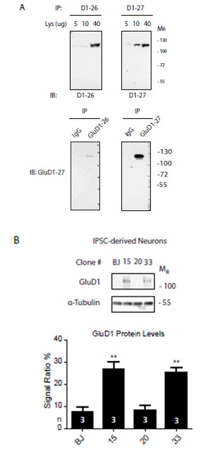
Figure 3.

(A). Anti-GluD1 antibody characterization. Different amounts of mouse brain lysate as well as immunoprecipitates (with mouse non-specific IgG as control) were loaded and probed for two different antibody batches (#26 and #27) to double-proof the identity of the recognized epitope. (B). GluD1 expression in iPSC-derived neurons. Each lane was loaded with 30 μg of total protein. Experiments were performed in triplicate (n=3). Immunosignals were detected using autoradiographic films, Glutamate-Delta-1 receptor levels were quantified using Photoshop CS3 software and normalized against alpha-tubulin levels (**p<0,01, student t test).
