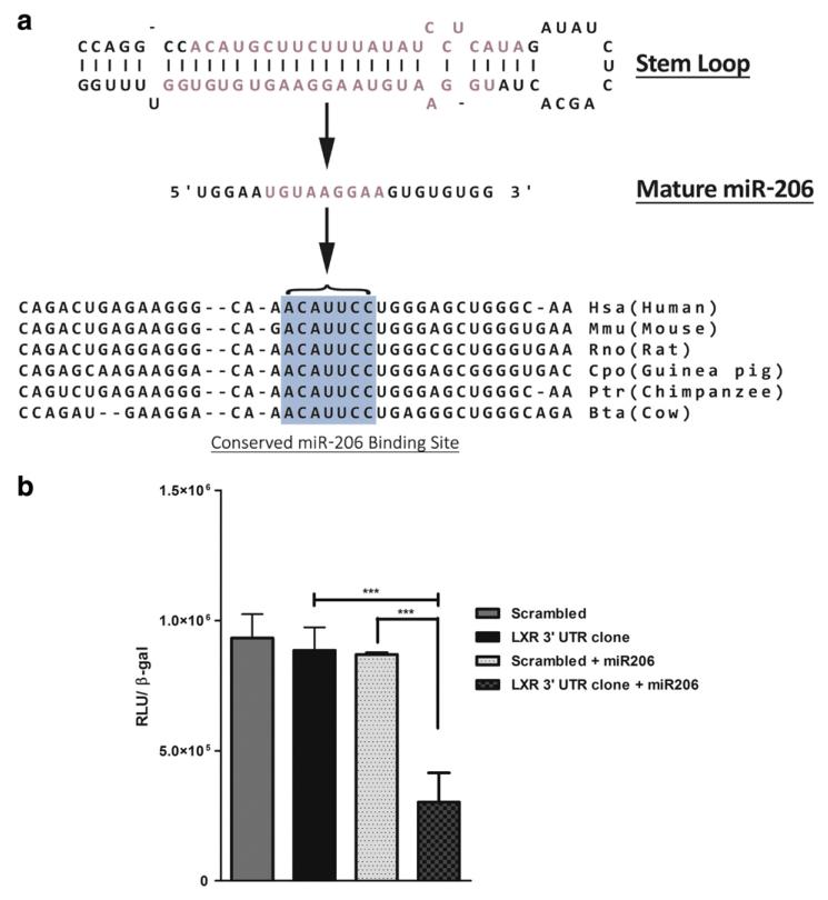
Fig. 1.
a) Conserved binding site for miR-206 on LXRα mRNA. Top: Stem loop structure of miR-206 from which the mature miR-206 is formed. The seed sequence of miR-206 that can putatively bind to the LXRα 3′UTR region is represented in pink and the conserved miR-206 binding site on LXRα mRNA is highlighted in blue. b) Luciferase reporter assay: Relative luciferase activity of the LXR 3′UTR construct and the 3′UTR scrambled construct. Mean values ± SEM from three separate experiments are as indicated;***p < 0.001.

