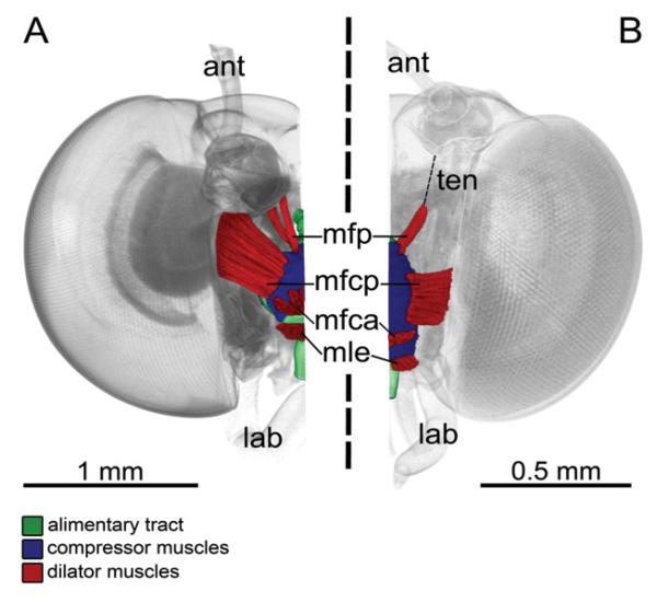
Figure 4. 3D reconstruction based on micro-CT images of the suction pump musculature of long-tongued (A) and short-tongued (B) Riodinidae, frontal view, proboscis removed.
A, Eurybia lycisca (long-tongued): right half of the head with alimentary tract, compressor muscles and dilator muscles [musculus frontoclypeo-pharyngealis (mfp), musculus frontoclypeo-cibarialis posterior (mfcp), musculus frontoclypeo-cibarialis anterior (mfca), musculus labro-epipharyngealis (mle)] of the suction pump. Compressors contract and reduce the suctorial cavity to swallow nectar into the oesophagus. Dilator muscles expand the suctorial cavity to produce a pressure gradient to take up nectar through the food canal of the straw-like proboscis (ant, antenna; lab, labial palpus). B, Sarota gyas (short-tongued): left half of the head, with alimentary tract, compressor muscles and dilator muscles of the suction pump. The dilator mfp is connected to its attachment site via a tendon (ten).

