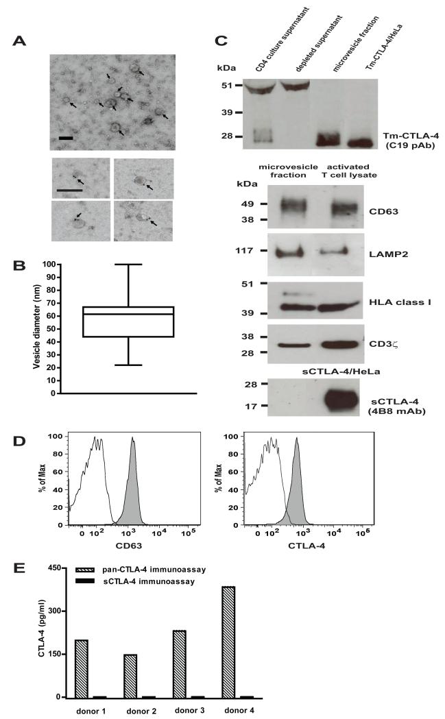
Figure 5.
Characterization of exosome-like vesicles secreted by activated CD4+ T cells. (A) Top panel: representative electron micrographs of microvesicles, as indicated by arrows, showing vesicles with low electron density as well as characteristic shape and size of exosomes (scale bar; 100 nm). Bottom panel: anti-CD63 immunogold labelling of CD4+ T cells-derived microvesicles. Arrows indicate immunogold-positive labelling (scale bar; 100 nm). (B) The size distribution of microvesicles is plotted as a box plot; the box span the 25thand 75th percentile, the horizontal line represents the median diameter and the whiskers mark the extremes of the distribution ( n = 24 microvesicles). (C) Top panel: Western blot analysis of activated CD4+ T cell supernatant before and after ultracentrifugation, microvesicle fraction isolated from the T cell supernatant and detergent lysates from Tm-CTLA-4/HeLa cells with anti-Tm-CTLA-4 C19 polyclonal Ab; bottom panels: Western blot analysis of microvesicle fraction isolated from the T cell supernatant with anti-CD63, anti-LAMP2, anti-HLA class I, anti-CD3ξ and anti-sCTLA-4 antibodies. CD4+ T cell or sCTLA-4/HeLa detergent lysates were used as positive controls. (D) Microvesicle fraction derived from activated CD4+ T cells were coated onto latex beads, surface stained with anti-CD63 and anti-CTLA-4 (BN13) (filled grey histogram) antibodies or the corresponding isotype controls (open histogram) and analyzed by flow cytometry. E) sCTLA-4 is not expressed in exosomal-like vesicles. The microvesicle fractions were isolated from supernatants of activated CD4+ T cells of 4 donors after 3 days of stimulation with anti-CD3/CD2 mAb and then tested in the pan-CTLA-4 and s-CTLA-4 immunoassays. All experiments are representative of at least three independent observations.

