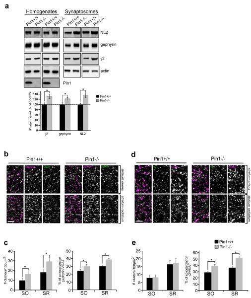
Figure 5. Synaptic enrichment of GABAARs is achieved in Pin1−/−.
(a) Representative immunoblots of NL2, gephyrin and γ2 subunit of GABAA receptor extracted from the hippocampus of Pin1+/+ and Pin1−/− mice (littermates) in two different sets of experiments. Total proteins from the homogenates and synaptosome suspension fractions were analysed by western blotting. Below: quantification of the indicated antigens extracted from hippocampal tissues of Pin1+/+ and Pin1−/− mice. All markers analyzed are enriched at inhibitory synapses. Western blot to actin was done as loading control. Pin1immunoblot indicates hyppocampus from Pin1+/+ and Pin1−/− (n=6 littermate pairs, mean values ± s.d, *p < 0.05, Student’s t-test) Full images of western blots are in Supplementary Fig.5. (b) Representative confocal micrographs of frontal brain sections showing segments of the str. radiatum (SR) and str. oriens (SO) of the CA1 region of the hippocampus from adult Pin1+/+ and Pin1−/− mice immunolabeled for gephyrin (magenta) and VGAT (green). Scale bar: 5μm. (c) Quantification of gephyrin punctum density (normalized to 100 μm2) and their percentage of colocalization with the presynaptic marker VGAT in both mouse genotypes. (d). Confocal micrographs as in (a) immunolabeled for GABAA receptor γ2 subunit (green) and VGAT (magenta). (e) Quantification of γ2 subunit punctum and their percentage of colocalization with VGAT in both mouse genotypes. The number of gephyrin, γ2, gephyrin and VGAT puncta was assessed in at least 8 sections for each genotypes (Pin1+/+ and Pin1−/−), by taking at least 4 images of strata radiatum and oriens of the CA1 region of each hippocampus in each set of experiments (n=3). Mean values ± s.d., *P < 0.05, Student’s t-test. Scale bar: 5μm.

