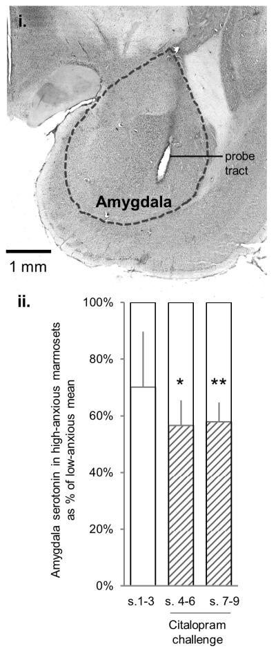
Figure 4. Amygdala microdialysis (in vivo extracellular serotonin levels).

(i) Coronal section through the amygdala illustrating the positioning of the microdialysis probe. (ii) Extracellular levels of amygdala serotonin in high-anxious marmosets expressed as % of the mean low-anxious levels, at baseline (white bars, samples 1-3, no citalopram) and following citalopram challenge (shaded bars, samples 4-6 and samples 7-9). *p < 0.05, **p < 0.01 compared to low-anxious mean. Although three out of five high-anxious animals also showed substantially reduced serotonin levels at baseline (39-52% of low-anxious mean), this did not reach statistical significance for the group (samples 1-3, t(4) = −1.53, p = 0.49).
