Figure 1.
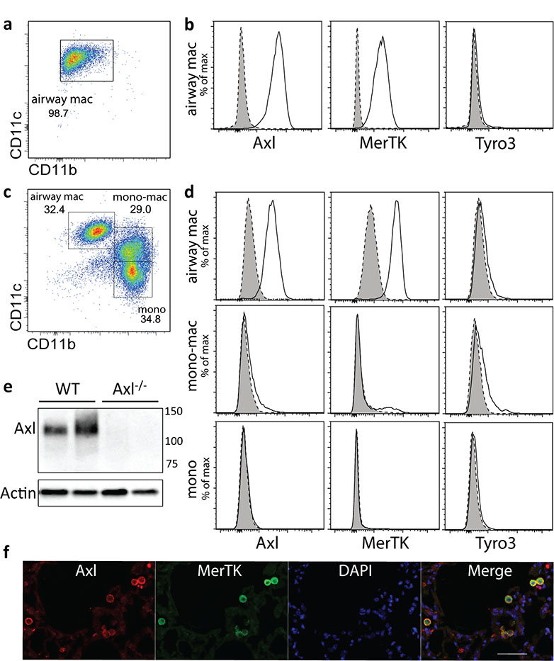
Axl is primarily expressed on airway macrophages in homeostasis
Flow cytometric analysis of F4/80 positive cells in (a) airway and (c) dissociated interstitial lung tissue by CD11b and CD11c expression. Airway macrophages (airway mac) are defined as CD11bloCD11chi; lung monocyte-macrophages (mono-mac) are defined as CD11bhiCD11cintermediate; lung monocytes (mono) are defined as CD11bhiCD11clo. (b and d) Flow cytometric analysis of Axl, MerTK and Tyro3 expression on airway macrophages, lung monocyte-macrophages, and lung monocytes. Specific staining, solid line/open. Isotype control, dotted line/shaded. (e) Western blot analysis of Axl protein expression by airway macrophages isolated from wild type (WT) or Axl−/− mice. Each lane represents lysates from individual mice. (f) Paraffin embedded lung sections from naïve mice stained with antibodies specific for Axl (red) or MerTK (green), and counter-stained with DAPI (blue). Scale bar: 50 μm. Data are representative of two independent experiments with four to five mice (all except f) or two independent experiments with two mice per group (f, immunohistochemistry).

