Fig. 4.
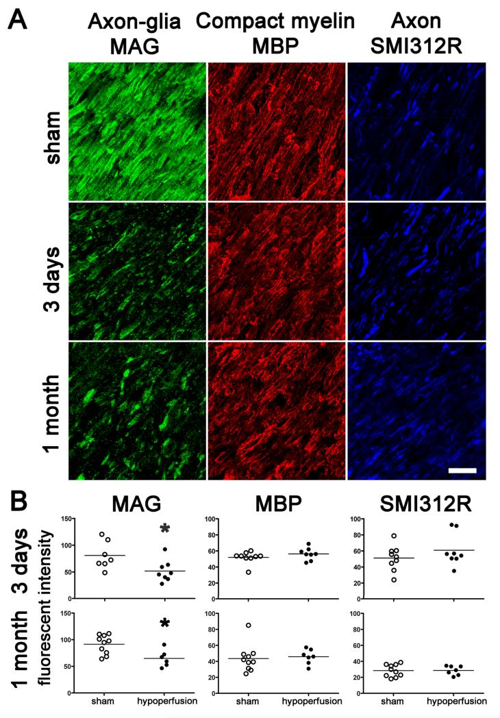
Axon-glial integrity is disrupted whereas myelin and axonal integrity remains intact. (A) Disruption of axon-glial integrity was defined as reduced and discontinuous granular accumulation of the MAG staining, in response to hypoperfusion as compared to shams. In contrast, the integrity of the myelin sheath (assessed by MBP) and integrity of axons (assessed by SMI312R) remains intact at 3 days and 1 month. Scale bar = 10 μm. (B) Measurement of the fluorescent intensity of MAG, MBP and SMI312R staining was conducted by confocal laser microscopy in the corpus callosum, internal capsule and optic tract in sham and hypoperfused groups at 3 days (sham, n = 9; hypoperfused n = 8) and 1 month (sham, n = 10; hypoperfused n = 7). There was a significant reduction in the intensity of MAG staining at both 3 days and at 1 month hypoperfusion as compared to shams. There were no alterations in MBP and SMI312R immunostaining in response to hypoperfusion at any time studied. *, P < 0.05 (Mann-Whitney, two-tailed).

