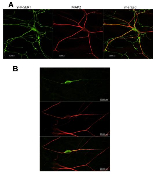
Figure 4.
YFP–SERT–607RI608–AA fails to reach axonal compartments. Rat dorsal raphe neurons were transfected with plasmids encoding YFP–SERT (A) or YFP–SERT–607RI608–AA (B) using Lipofectamine 2000. After 48 h, the neurons were fixed and stained for MAP-2. MAP-2 staining was detected using an Alexa Fluor 568-conjugated secondary antibody. Images were captured by confocal microscopy. YFP-tagged SERT was found in the MAP-2-negative compartment in 24 of 24 examined neurons; YFP-tagged SERT–607RI608–AA was confined to the MAP-2-positive compartment in 20 of 21 examined neurons. Data are from three independent experiments (i.e., 4 individual preparations of rat dorsal raphe neurons that were independently transfected in parallel with a plasmid encoding either YFP–SERT or YFP–SERT–607RI608–AA). Scale bars, 20 μm.

