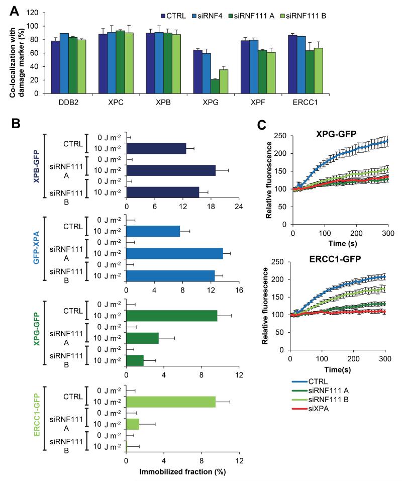
Figure 3. RNF111 is required for binding of XPG and XPF/ERCC1 to the NER complex.
(A) U2OS cells expressing CPD-photolyase-mCherry were transfected with the indicated siRNA’s three days before the immunofluorescence experiment. Cells were local UV-irradiated (60 J m−2) and immunostained for the indicated proteins 30 min later. The percentage of co-localization with the damage marker CPD-photolyase-mCherry at LUD is plotted in the graph (n>50cells containing a LUD were scored in at least three independent experiments; error bars are the mean ± SD). (B) The immobilized fraction of XPB-GFP, GFP-XPA, XPG-GFP and ERCC1-GFP as determined by FRAP analysis in mock or UV-treated (10 J m−2) cells upon transfection with the indicated siRNA’s (n>32 cells from at least 2 independent experiments; error bars are the mean ± 2* SEM). (C) Cells stably expressing XPG-GFP and ERCC1-GFP transfected with the indicated siRNA’s were locally irradiated using a 266 nm UV-C laser. GFP fluorescence intensity at LUD was monitored for 6 min, with 10 s intervals and normalized to pre-damage values. (n=24 cells from three independent experiments; error bars are the mean ± SEM).

