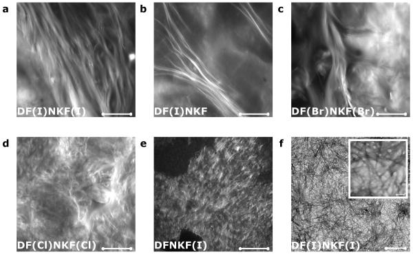
Figure 3. Fibrillar morphologies of halogenated hydrogels.
a-e, Confocal microscopy images of peptide hydrogels at a concentration of 15 mM upon aging 48 hours at r.t. and after staining with Rhodamine B (scale bars, 100 μm). a, b, and c show bundles of long twisted fibrils belonging to hydrogels of peptides DF(I)NKF(I), DF(I)NKF, and DF(Br)NKF(Br), respectively. d and e show much smaller fibrils of hydrogels of peptides DF(Cl)NKF(Cl) and DFNKF(I), which intertwine and form a matrix-type structure. f, TEM image of the dried hydrogel of peptide DF(I)NKF(I) showing an intertwined network of long fibrils (scale bars, 0.5 μm and 5 nm).

