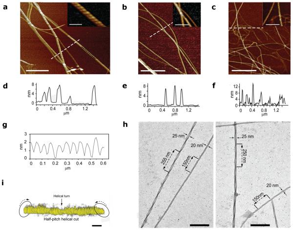
Figure 4. Nanofibrillation of the halogenated pentapeptides.
AFM image close-up insets of a, peptide DF(I)NKF(I), b, peptide DF(I)NKF, and c, peptide DF(Br)NKF(Br) evaporated on mica substrates after 9 days incubation in aqueous solutions (scale bars, 1 μm). d-f, Height profiles of fibrils from a, b, and c (cross-section lines highlighted in white). g, Cross-sectional analysis on the top of a fibril segment in a showing a helicoidal profile along the longitudinal direction of the fibrils (highlighted in black). h, Cryo-TEM images of the dried hydrogel of DF(I)NKF, showing a network of long fibrils (scale bars, 200 nm). i, In-situ electron tomography of half-pitch helicoidal peptide DF(I)NKF(I) vitrified from aqueous solution. The twisted fibril morphology is highlighted by arrows. Electron tomography reconstructions were collected upon tilting of samples up to ± 69° (scale bars, 25 nm).

