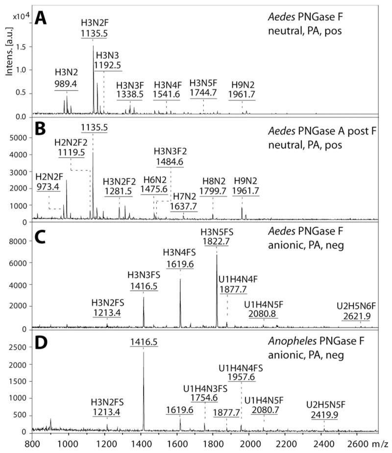
Figure 1. Mass spectrometric analysis of neutral and anionic mosquito N-glycan pools.
Released N-glycans were enriched in neutral and anionic pools prior to pyridylamino-labelling and MALDI-TOF/TOF MS. The spectra shown are those in positive (A, B) and negative ion mode (C, D) of the N-glycan pools released with PNGase F (A, C, D) or PNGase A post PNGase F (B) from Aedes aegypti (A-C) and Anopheles gambiae (D). Abbreviations for annotated [M+H]+ and [M−H]− ions are: H Hexose, N N-acetylhexosamine, F Fucose, S Sulphate and U Glucuronic Acid. The positive ion mode spectra of the neutral Anopheles PNGase F and A pools are not shown due to their similarity to the Aedes; the differences between the two species, limited to the lower intensity glycans, were only resolved after HPLC fractionation.

