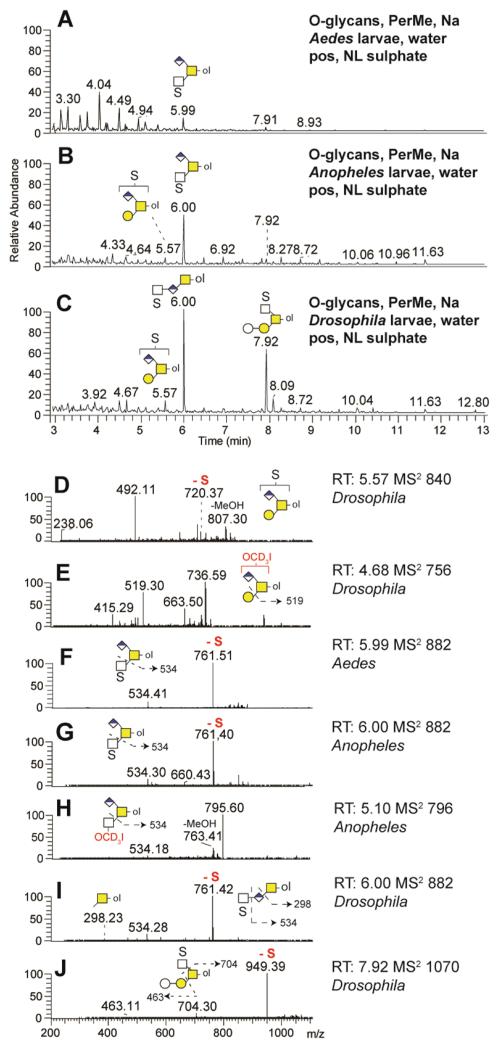
Figure 13. Sulphated O-glycans analyses by NSI-MS.
Sulphated O-glycans were released by reductive β-elimination, permethylated, enriched in the water phase and analysed by NSI-MS in positive and negative ion mode. In automated positive ion mode TIM, spectra were filtered for neutral loss (NL) of sodiated sulphate moieties (120 Da) in Aedes (A), Anopheles (B) and Drosophila (C) larvae preparations. Three singly charged sulphated O-glycans were detected, two O-glycans carrying both sulphate and glucuronic acid modifications (m/z 840 and 882) and one sulphated O-glycan (m/z 1070). The automated TIM MS2 spectra of selected O-glycan structures found in Anopheles (G, H), Drosophila (D, E, I, J) and Aedes (F) larvae are presented together with potential fragmentation schemes.

