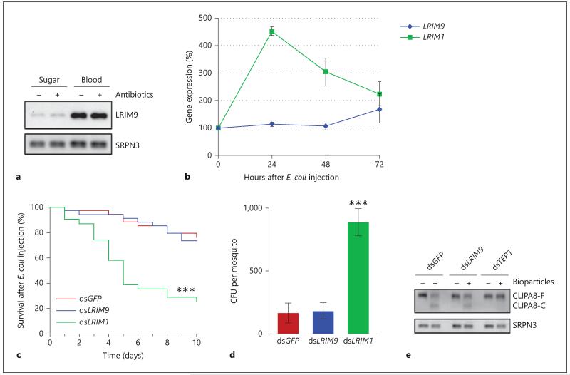
Fig. 4.
LRIM9 is not regulated by bacteria or involved in antibacterial defense. a Newly emerged mosquitoes were fed either sterile sugar solution or a cocktail of antibiotics dissolved in sugar. After 4 days of treatment, some mosquitoes were allowed to feed on uninfected murine blood whilst others were kept on sugar. After 24 h, hemolymph was collected and analyzed by nonreducing Western blot, probing with LRIM9 and SRPN3 antibodies. b RNA was extracted from mosquitoes 24, 48 and 72 h after E. coli injection. LRIM9 and LRIM1 expression was determined by qRT-PCR, normalizing to ribosomal S7. Expression was normalized to injection of sterile PBS at each time point and calculated relative to uninjected (0 h) mosquitoes. The mean of 2 independent experiments is shown with standard error bars. c Mosquitoes were injected with dsGFP, dsLRIM9 and dsLRIM1, blood fed after 3 days and innoculated with live E. coli 24 h later. Mosquito survival was monitored daily for 10 days. Survival was compared to dsGFP using the Kaplan-Meier log-rank test (*** p < 0.0001). d Mosquitoes were injected with dsGFP, dsLRIM9 and dsLRIM1 and after 4 days injected with live ampicillin-resistant E. coli. After 24 h, batches of 10 mosquitoes were surface sterilized, washed and homogenized. The homogenate was plated onto ampicillin LB agar and colony-forming units (CFU) were counted after overnight incubation at 37°C. Mean CFU per mosquito from 3 independent experiments is shown with standard error bars. dsLRIM9 and dsLRIM1 were compared with dsGFP using meta-analysis (*** p < 0.0001). e Four days after injection of dsGFP, dsLRIM9 and dsTEP1, hemolymph was collected from half of these mosquitoes. The other half were injected with Staphylococcus aureus bioparticles, and hemolymph was collected 2 h after challenge. Hemolymph was analyzed by reducing Western blot and probed with antibodies against CLIPA8 and SRPN3 (loading control).

