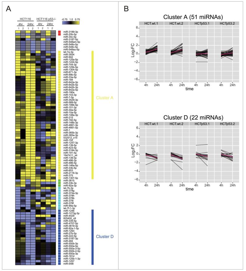
Figure 9.
TP53 shapes genome-wide patterns of human miRNA expression during the DNA damage response. (A) Shows the heat map of expression patterns for 93 DDR-responsive miRNAs in HCT116 and HCT116TP53−/− cells at 4 h and 24 h after DNA damage, compared to their expression at 0 h. Two technical replicates (1 and 2) were analyzed for each condition. Each row represents a specific miRNA species. miRNAs were clustered by the pattern of the expression into clusters as in Fig. 5. (B) Shows changes in the expression of cluster A (miRNAs induced during the DDR) and cluster D (miRNAs repressed during the DDR). Note that the induction of cluster A miRNAs is lost in HCT116TP53−/− cells, but not the repression of those in cluster D.

