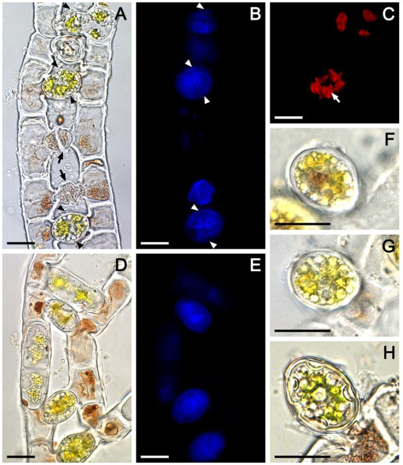
Figure 1.
Light microscopic (A, D, F-H) and autofluorescence (B, C, E) images of consequent conjugation stages in Zygogonium ericetorum: (A-C) same conjugating filaments showing healthy united gametes and zygospores, and abnormal gametangia with incomplete conjugation (black arrows), (B) blue autofluorescence of zygospore cell wall compounds, arrowheads show the rupture along the contact line between the two corresponding conjugation tubes enclosing healthy united gametes and zygospores, (C) red autofluorescence of chloroplasts in gametes and zygospore, white arrow shows the pyrenoid, (D, E) same conjugating filaments in early conjugation stage, note that the united gametes and zygospores are separated from the purple-colored cytoplasmic residue left in gametangia by wall, (F, G) zygospores with thick smooth multilayered colorless spore wall detached from one of the conjugating filament, (H) completely developed zygospore with thick smooth multilayered yellowish spore wall detached from one of the conjugating filament. Scale bars: 20 μm.

