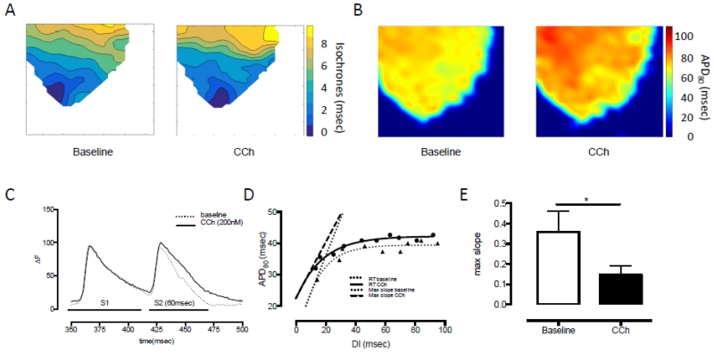
Figure 2. Carbamylcholine perfusion and effects on ventricular electrophysiology.
A. Conduction velocity during CCh perfusion and apical pacing remains unchanged while there is B. a significant increase in median APD80 as determined by optical mapping of the anterior wall of the left ventricle. C. CCh perfusion results in prolongation of the APD in response to a closely coupled extra-stimulus following a drive train compared to baseline conditions (ΔF: fractional change in RH237 fluorescence). D/E. This results in a significant (*p<0.05) flattening of the electrical restitution slope (RT: restitution, max slope: steepest part of the restitution curve).

