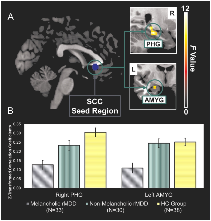
Figure 1.
a) The network of regions demonstrating resting-state functional disconnection with the subgenual cingulate seed region in the remitted melancholic MDD patients when compared to the remitted non-melancholic MDD and HC groups. Whole-brain images were cropped and displayed at an uncorrected voxel-level threshold of p<0.001. b) Bar plots showing group differences in average Z-transformed correlation coefficients and standard errors for the right parahippocampal gyrus and left amygdala clusters. AMYG, amygdala; HC, healthy control; L, left; MDD, major depressive disorder; PHG, parahippocampal gyrus; R, right; SCC, subgenual cingulate cortex.

