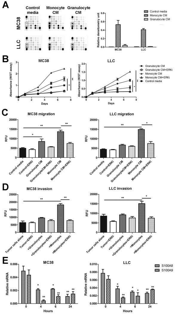
Figure 3.
Effects of ERK signaling in MC38 and LLC tumor cells. (A) MC38 and LLC cells were cultured in monocyte/macrophage- and granulocyte-conditioned media and activation of ERK signaling pathway (boxed area) assessed using the PathScan Intracellular Signaling Array Kit by measuring densitometry values. Three independent experiments were performed, representative image shown. MC38 and LLC (B) proliferation, (C) migration and (D) invasion in response to monocytes/macrophages or granulocytes were tested in the presence of the ERK inhibitor U0126 (10 μM; +ERKi) using the WST or Cytoselect assays. Three independent experiments were performed, *p<0.05, **p<0.01. (E) Expression of S100a8 and S100a9 mRNA in MC38 and LLC cells cultured in monocyte/macrophage-conditioned medium were assessed by qPCR after treatment with 10 μM ERK inhibitor U0126 for the indicated times. Three independent experiments were performed, *p<0.05, **p<0.01 compared to time 0 h.

