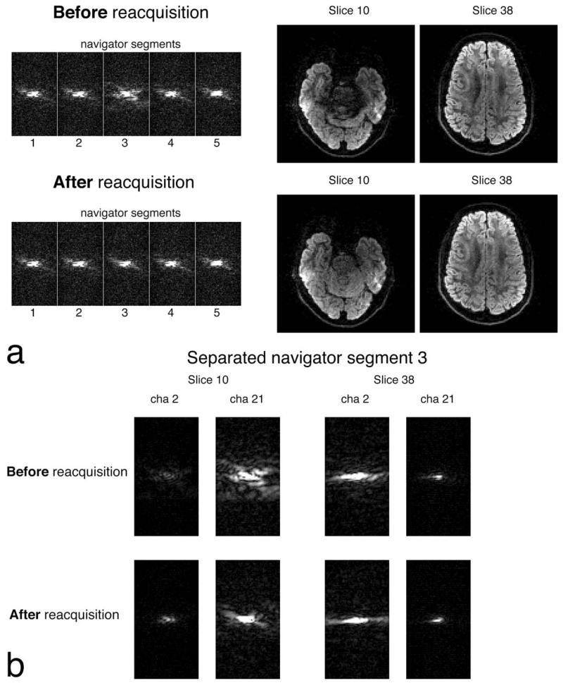
FIG. 3.
Navigator-based reacquisition with two simultaneously excited slices (labelled with their anatomical slice number) with b = 1000 s/mm2 diffusion-weighting. a: Before reacquisition, corruption in segment 3 is indicated by the dispersed k-space in the navigator, which contains combined data from the two slices. When segment 3 is reacquired, artifacts are removed from the images. b: Single-channel maps (all at the same scale) of the unaliased k-space from navigator segment 3. Two channels are chosen which are close to slice 10 (channel 21) and 38 (channel 2) to demonstrate the variation in signal.

