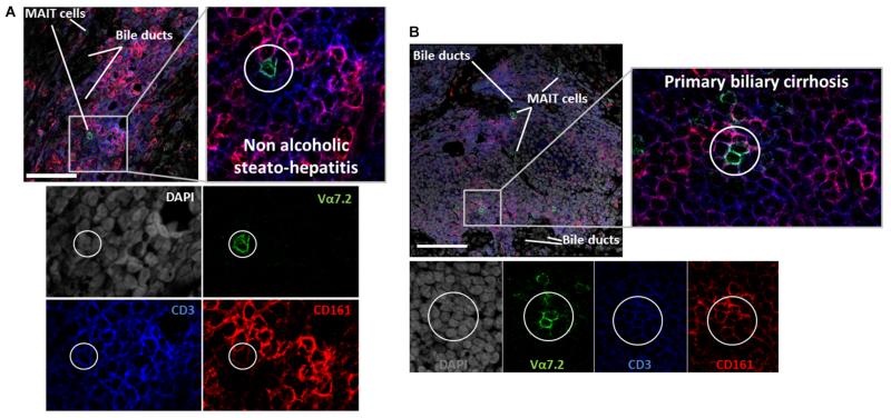
Figure 2. CD3+CD161+Va7.2+ cells reside close to bile ducts in portal tracts.
Representative confocal immunofluorescence staining for CD3, CD161 and Va7.2 on frozen sections from explanted human livers diagnosed with Non Alcoholic Steato-Hepatitis (A) and Primary Biliary Cirrhosis (B). DAPI nuclear stain reveals liver architecture indicating sites of bile ducts. Images are representative of staining of four different diseased livers, scale bar shows 100μm.

