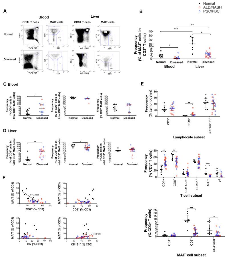
Figure 3. Flow cytometric analysis of MAIT cell frequencies in chronic liver disease and their correlations with other immune subsets.
Representative FACS plots (A) and summary frequency data for total CD3+ MAIT (B) and CD4+, CD8+ and CD4−CD8− (DN) MAIT subsets in normal and diseased blood (C) and liver (D). (E) Frequencies of intrahepatic MAIT and other immune cells. (F) Correlation of CD3+ MAIT cell frequencies with total CD4+, CD8+, DN and CD161+ T cell frequencies in normal and diseased livers. Data are median ± interquartile range. *=p<0.05; **=p<0.01; ***=p<0.001 by Mann Whitney test (B-E) or Spearman’s rank correlation (F).

