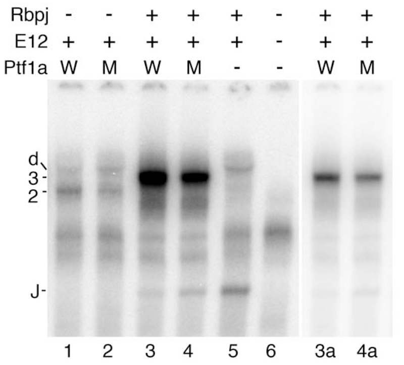
Figure 3. The DNA binding of wild type PTF1A and PTF1A-P191T as part of the PTF1A:E12 dimer or PTF1A:E12:Rbpj trimer in EMSA.
The relative abilities of wild type and p.P191T mutant Ptf1a to form complexes with E12 and Rbpj on the PTF1 binding site of the Rbpjl gene was tested in vitro. W, Ptf1a wild type; M, p.P191T mutant; 2, Ptf1a:E12 dimer; 3, Ptf1a:E12:Rbpj trimer; d, E12 homodimer; J, Rbpj monomer. Lanes 3a and 4a show lanes 3 and 4 with an expanded gray scale such that the PTF1 trimeric complex bands are not saturated. Ptf1a alone does not bind DNA.

