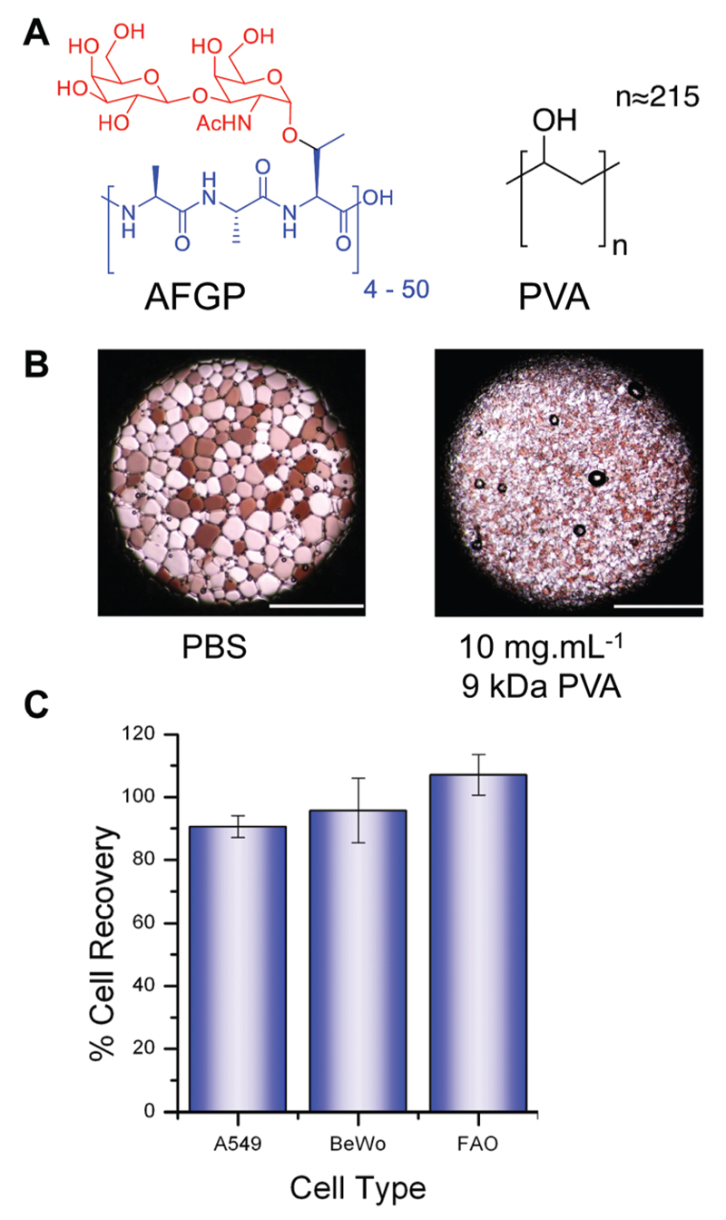
Fig. 1.
PVA structure, IRI activity and cytotoxicity. (A) Structure of AF(G)P and PVA; (B) micrographs of ice crystals annealed for 30 minutes at −6 °C showing inhibitory effect of PVA; (C) cytotoxicity testing of 25 mg mL-1 PVA (9 kDa) after 4 hour incubation. Cell metabolic activity measured by MTT (A549) and resazurin (BeWo and FAO) assays. Error bars represent ± standard deviation from at least 3 repeats.

