Figure 3.
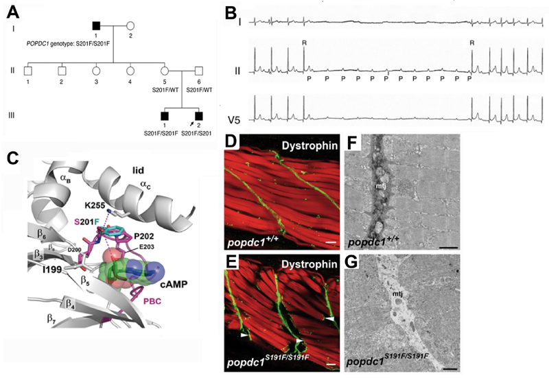
The POPDC1S201F missense mutation causes cardiac arrhythmia and muscular dystrophy. (A) Pedigree of an Italian family homozygous for the POPDC1S201F mutation. The index patient (III-2, arrow) and his brother (III-1) suffer from AV block, while the grandfather (I-1) has both, AV block and LGMD. (B) Holter electrocardiogram of the index patient displaying an episode of a paroxysmal AV block. (C) 3D model of the Popeye domain. Only the phosphate-binding cassette (PBC) is shown. Two hydrogen bonds are formed by S201, but these are predicted to be lost when the S201 residue is replaced by a phenylalanine (turquoise) affecting the ligand-binding affinity of the Popeye domain. (D-G) are sections through the zebrafish tail musculature. (D,E) Confocal analysis of actin (phalloidin staining, red channel) and Dystrophin (green channel) in trunk skeletal muscle of (D) WT (popdc1+/+) and (E) homozygous zebrafish mutant (popdc1S191F/S191F) at 5 dpf. Homozygous mutants develop lesions in skeletal muscle. Muscle fibers detach from the MTJ and retract. Arrowheads indicate the presence of dystrophin immunoreactivity at the end of the ruptured fibers, suggesting that the fibers have lost connection to the MTJ but probably remain physically intact. (F,G) TEM analysis of trunk skeletal muscle of (F) WT and (G) homozygous popdc1S191F mutant zebrafish embryos at 5 dpf. The MTJ in the homozygous mutant is characterized by a lack of electron-dense material. Panels A–G are reproduced from [12] with permission.

