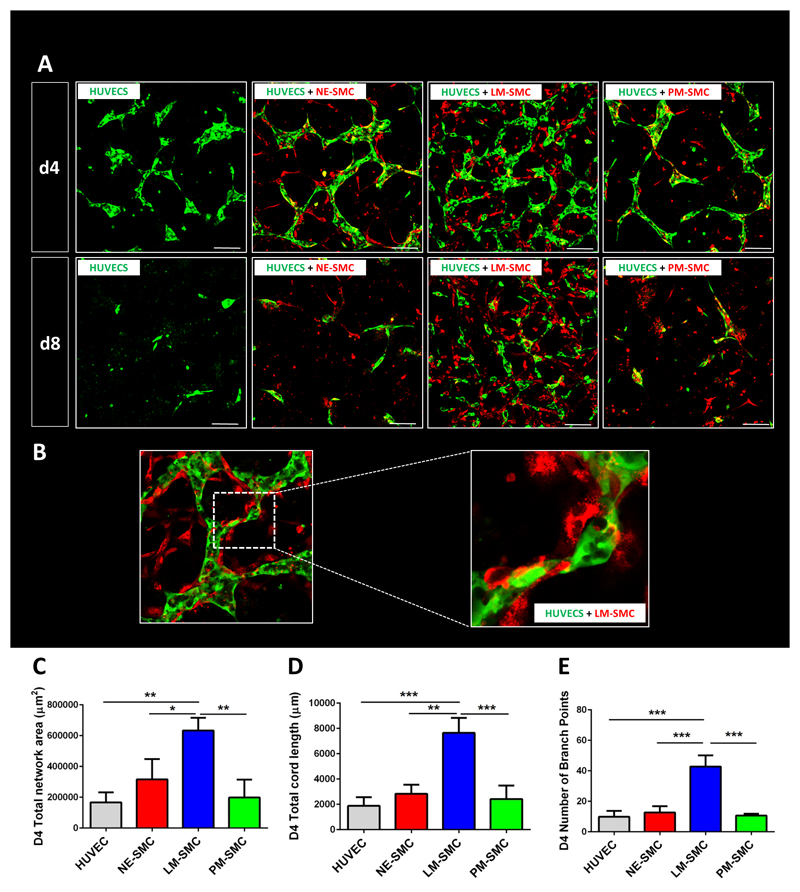
Fig. 2. LM-derived SMCs best support HUVEC network formation in a 3D co-culture model.
(A) Confocal images of HUVEC (green) co-cultures with the three respective SMC types (red) on day 4 and day 8 of the co-culture protocol. (B) SMCs provide physical support to developing networks, wrapping around endothelial network. (C) Quantification of total network area in μm2. (D) Quantification of total cord length. (E) Quantification of number of branch points. Quantitative data is shown for day 4 of the co-culture (*p<0,05, **p<0,01, ***p<0,001, n=3 independent biological triplicates, scale bars= 100 μm).

