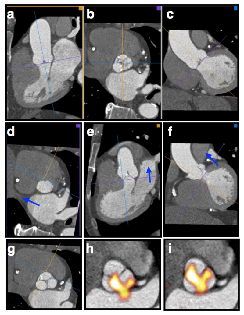
Figure 1. Creation of Co-registered En Face Short-Axis PET/CT Images of the Aortic Valve.
Creation of planar en-face valve images using CT angiography was achieved as follows. First, the CT angiogram is reorientated to get into the approximate plane of the aortic valve by lining up the axial cross hair (purple in this example) using the images in the coronal (a) and sagittal planes (c). This creates an approximate cross sectional image of the aortic valve in the axial frame (b). Scrolling down in the axial frame, the center of the crosshairs is then placed over the exact point at which the right coronary cusp disappears, identifying the base of that leaflet (d). Similarly the base of the non-coronary cusp is identified and orthogonal planes adjusted so that the purple plane goes through the base of both these two cusps (d). Finally the base of the left coronary cusp is found by rotation of the axial cross hairs so that first the cusp comes into view. The image is then slowly rotated in the opposite direction until the point where the leaflet first disappears (the base) is again found (f). This produces an en face image of the valve aligned with the base of all three leaflets (g). Adjacent 3-mm slices are then created in that plane and used for subsequent assessment. These slices are fused with the 18F-Fluoride PET images (h) and careful co-registration performed in 3-dimensions to ensure accurate alignment between the PET and CT images (i).

