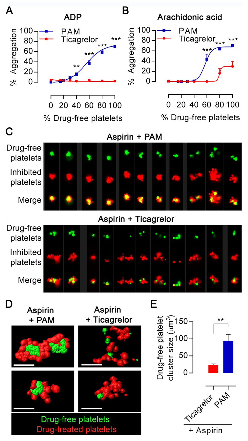
Figure 1. Ticagrelor reduces aggregation and prevents formation of drug-free platelet cores during aggregate formation in vitro.
PRP derived from blood pre-incubated with aspirin (30µM) and PAM (3µM) or ticagrelor (1.35µM) was mixed in a range of proportions with PRP from blood pre-incubated with respective vehicles or with ticagrelor (1.35µM) to reflect mid-dose t=6hour levels. Aggregation in response to (A) ADP 20µM or (B) AA 1mM was determined by LTA. Data presented as mean±SEM and compared by two-way ANOVA (n=4, **p<0.01, ***p<0.001). (C) Multiple images captured by ImageStreamX of aggregates (mixtures of 85% aspirin+PAM-pretreated platelets or aspirin+ticagrelor pretreated platelets plus 15% uninhibited platelets). Each panel contains columns with following image sets: drug-free (green), inhibited platelets (red), merged image. (D) Representative confocal images of aggregates (left) formed from mixtures comprising 85% aspirin+PAM-pretreated platelets or aspirin+ticagrelor-pretreated (green) and 15% uninhibited platelets (red). (E) Images were analysed for size of the uninhibited platelet particles. Data is presented as mean±SEM and compared by t-test (n=4, **p<0.01).

