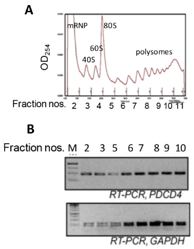
Figure 2. Ribosomal fractions from MCF7 cells were analysed by 10-50% sucrose density gradient fractionation.
A. Ribosomal RNA content, measured at 254 nm, is plotted against fraction numbers (top panel). B. RNA isolated from selected fractions was analyzed by semiquantative RT-PCR using PDCD4 and GAPDH primers (bottom panel).

