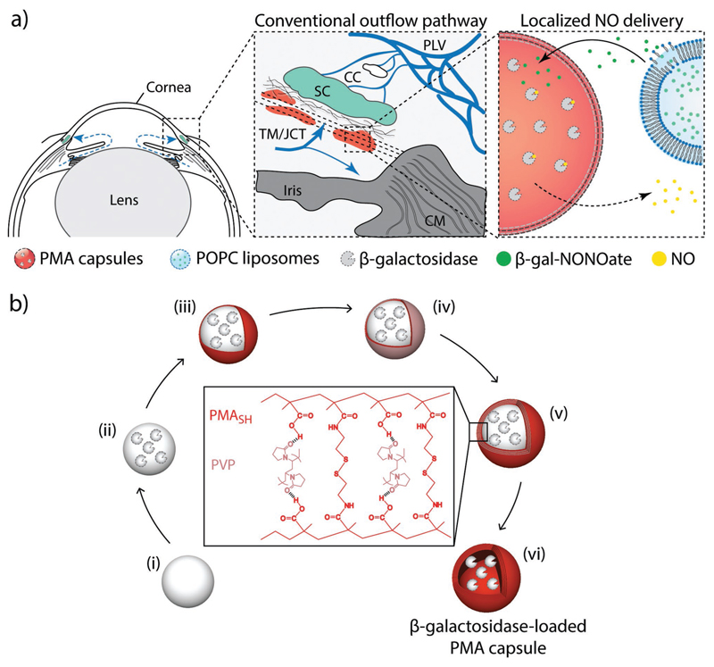
Scheme 1.
a) Schematic illustration of localized delivery of nitric oxide (NO) to the conventional outflow pathway via enzyme biocatalysis. Left: Anterior segment of eye showing the direction of aqueous humor flow in blue. Center: Enlargement of the iridocorneal angle (boxed region in left panel) showing the conventional outflow pathway. Right: Schematic of localized NO delivery within the trabecular meshwork (TM) near Schlemm's canal (SC). β-Galactosidase is encapsulated in poly(methacrylic acid) (PMA) capsules and enmeshed within the TM. Liposomes containing NO donors (β-gal-NONOate) are delivered to the outflow pathway. Upon liposome degradation, NO donors are slowly released at the outflow resistance sites and enzymatic activity of β-galactosidase results in local delivery of active therapeutic NO at the outflow resistance sites, achieving a targeted on-site NO delivery to the conventional outflow pathway. CC: collector channels, CM: ciliary muscle, JCT: juxtacanalicular connective tissue, and PLV: perilimbal vessels. b) Schematic illustration of assembly of β-galactosidase-loaded PMA capsules via layer-by-layer technique. i) Aminated silica particle template is coated with ii) β-galactosidase, followed by sequential deposition of iii) thiol-functionalized PMA (PMASH) and iv) poly(N-vinylpyrrolidone) (PVP) via hydrogen bonding. v) Once four bilayers of PMASH/PVP are deposited, the thiol groups of PMASH are oxidized into bridging disulfide linkages. vi) Removal of the sacrificial particle template results in (bio)degradable disulfide-cross-linked β-galactosidase-loaded PMA capsule.

