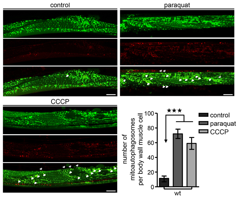
Figure 3. Monitor mitoautophagosomes formation in vivo.
Transgenic nematodes co-expressing a mitochondria-targeted GFP (mtGFP) in body wall muscle cells together with the autophagosomal protein LGG-1 fused with DsRed, were treated with paraquat or CCCP. Mitophagy stimulation is signified by co-localization of GFP and DsRed signals (for each group of images mitochondria are shown in green on top, autophagosomes in red below, with a merged image at the bottom). Increased number of mitoautophagosomes upon oxidatve and mitochondrial stress (n = 50; ***P < 0.001; one-way ANOVA). Size bars denote 20 μm. Images were acquired using a 40x objective lens. Error bars denote SEM values.

