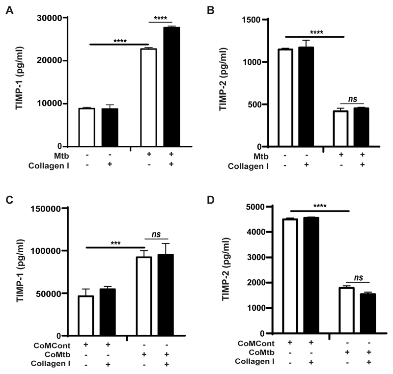
Figure 2. TIMP-1 and -2 secretion by Mtb-infected and CoMtb-stimulated monocytes adherent to type I collagen.
Monocytes adherent to type I collagen, fibronectin or in the absence of matrix were either infected with Mtb (MOI=1) or stimulated with CoMtb (1:5 dilution). Supernatants were collected at 24h post-infection and analyzed for secreted concentrations of: (A) TIMP-1and (B) TIMP-2 with Mtb-infected monocytes, and (C) TIMP-1 and (D) TIMP-2 secretion of CoMtb-stimulated monocytes. CoMCont was used as control of CoMtb. Bars show mean ±s.d. and figures are representative of 3 independent experiments performed in triplicate. ***p<0.001; ****p<0.001; ns- not significant.

