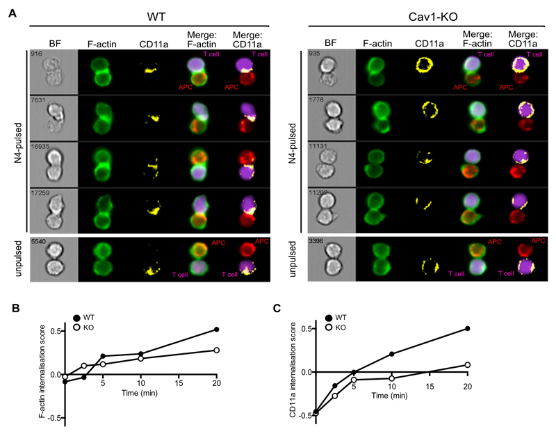
Figure 3. Impaired recruitment of F-actin and CD11a to the IS in the absence of Cav1. Imagestream analysis assessing conjugate formation of Cav1-WT or Cav1-KO OT-1 CD8 T cells with N4-pulsed APC for the indicated times (0-20 min). Conjugates were fixed, permeabilised and stained for BODIPY-Fl (F-actin, green) and CD11a (yellow).
(A) The 4 rows show representative cells at 20 min, with distribution of BODIPY-Fl and CD11a within the synapse formed between T cells (purple) and APC (red) shown in Merge columns 4 and 5 respectively. Graphs of the internalization method, previously described in (35), for Cav1-WT and Cav1-KO conjugates stained with (B) BODIPY-Fl and (C) CD11a are representative of 3 independent experiments.

