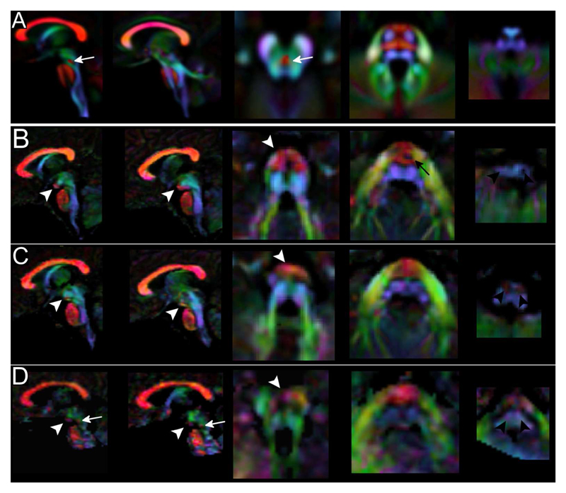
Figure 2. Color-coded DTI maps.
On color-coded maps patients 1 (Row B), 2 (Row C) and 3 (Row D) show an altered organization of white matter tracts if compared to a template of normal subjects (Row A). The anterior bulging of the mesencephalon corresponds to an area of transverse-oriented diffusivity located anteriorly in the interpeduncular fossa (white arrowheads). CST in the pons are thinned (black arrow) or non-clearly recognizable and transverse pontine fibers appear as a unique bundle displaced in the anterior part of the pons. In the medulla, CST and lemnisci are hypoplastic/atrophic and olives reduced in volume. The decussation of SCP (white arrow in the normal template) is absent in patient 1 and 2 and markedly thinned in patient 3 (white arrow in D). [Color legend: red, green and blue represent areas of respectively transverse, antero-posterior and caudo-cranial orientation of diffusivity and white matter]

