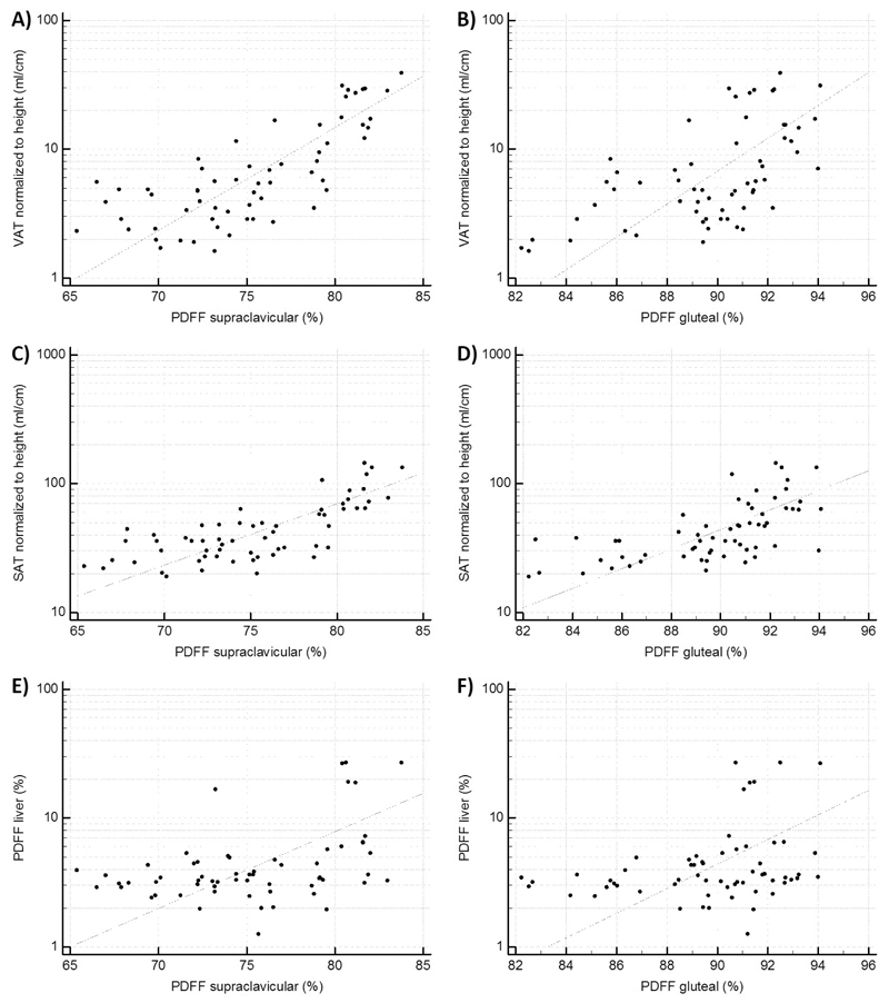
Figure 2.
Correlation analysis plots. A): Scatter plot of the correlation analysis of PDFF supraclavicular and VAT (r=0.76, p<0.0001). B): Scatter plot of the correlation analysis of PDFF gluteal and VAT (r=0.59, p<0.0001). C): Scatter plot of the correlation analysis of PDFF supraclavicular and SAT (r=0.73, p<0.0001). D): Scatter plot of the correlation analysis of PDFF gluteal and SAT (r=0.63, p<0.0001). E): Scatter plot of the correlation analysis of PDFF supraclavicular and PDFF liver (r=0.42, p=0.0008). F): Scatter plot of the correlation analysis of PDFF gluteal and PDFF liver (r=0.30, p=0.02). PDFF: proton density fat fraction, SAT: subcutaneous adipose tissue, VAT: visceral adipose tissue.

