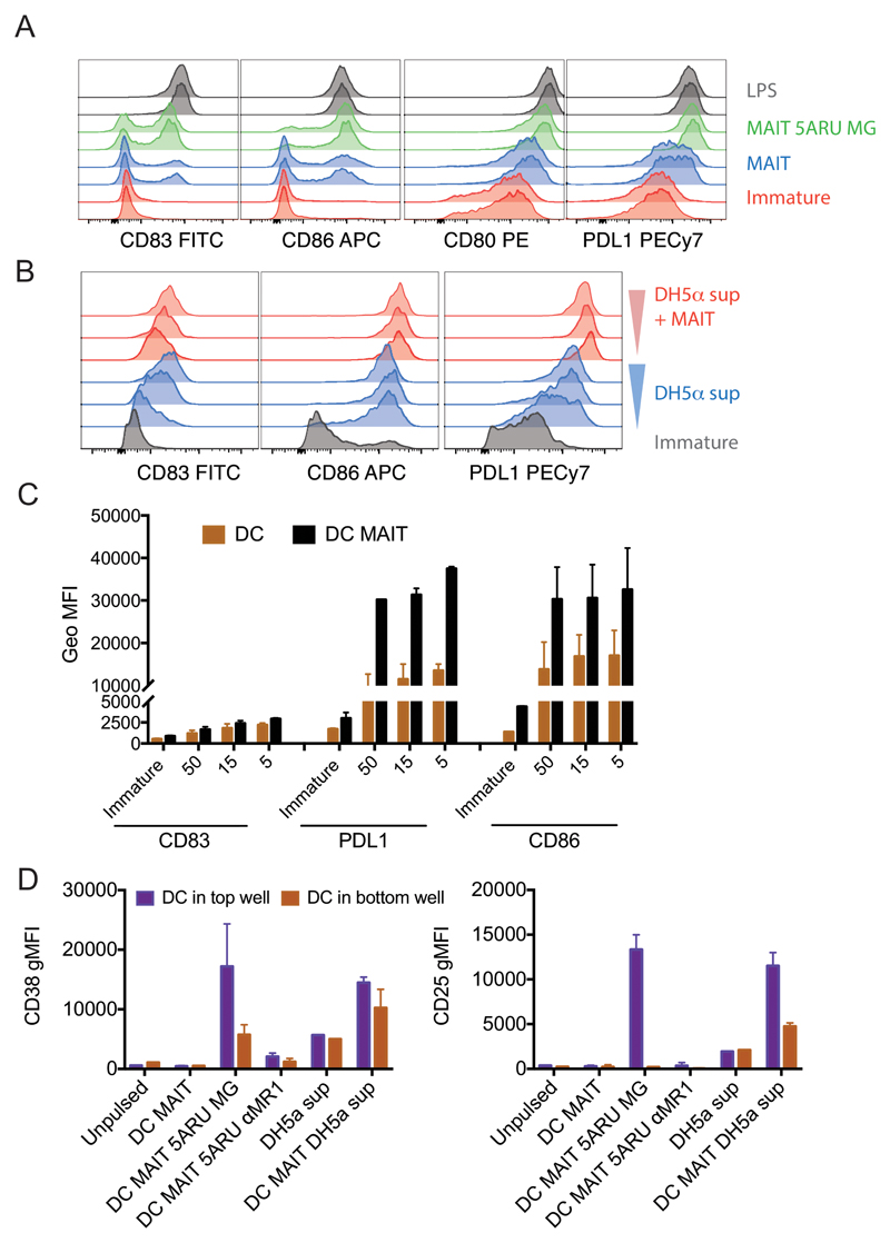
Figure 2.
Human MAIT cells induce DC maturation. A) Immature DC (red histograms) were incubated with allogeneic MAIT cells in the presence (green histograms) or absence (blue histograms) of 5-A-RU/MG. LPS (grey histograms) was used as positive controls. Staining profile of duplicate wells is shown. B) Immature DC (grey histograms) were incubated with three concentrations of DH5-α supernatant in the presence (red histograms) or absence (blue histograms) of autologous MAIT cells. Depicted are expression levels of CD83, CD86, CD80 and PDL1 as detected by flow cytometry after 36 hours. One representative experiment of four; data from two donors, one autologous and one allogeneic to the DC are tabulated in panel C, where 50, 15 and 5 on the x-axis refer to different amounts of DH5-α supernatant. D) Expression of CD38 and CD25 (geometric mean+/- SD of two donors) on DC harvested from the top and bottom wells of a transwell assay. When indicated, allogeneic MAIT cells were added in the top wells, in the presence or absence of 5-A-RU/MG or supernatant of DH5-α and anti MR1 blocking antibodies.

