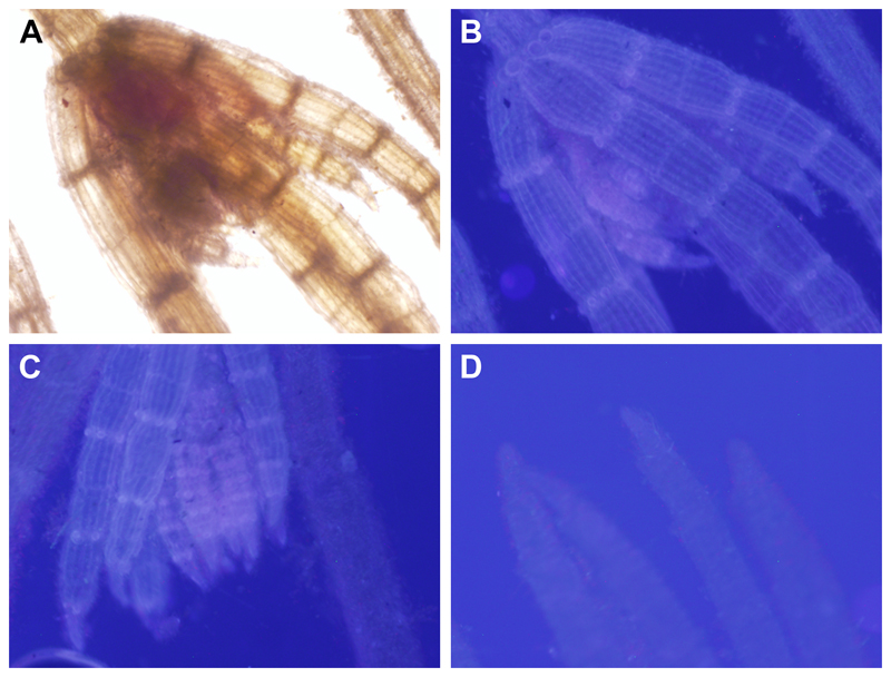
Fig. 4. Fluorescence microscopy images showing integration of the fluorescent acceptor substrate XyGO-SR into Chara sp. cell walls.
After incubation in XyGO-SRs, Chara was washed in culture media and viewed using the DAPI channel of an epifluorescence microscope at ×40 magnification. (A) Bright-field image. (B, C) Incorporation of XyGO-SRs in all cell walls. The walls of the stipulodes, and the cells at towards the tip of the main axis and branchlets appeared to have incorporated the most XyGO-SRs and fluoresced the most strongly. (D) Control in which Chara sp. was incubated in non-fluorescent XyGOs.

