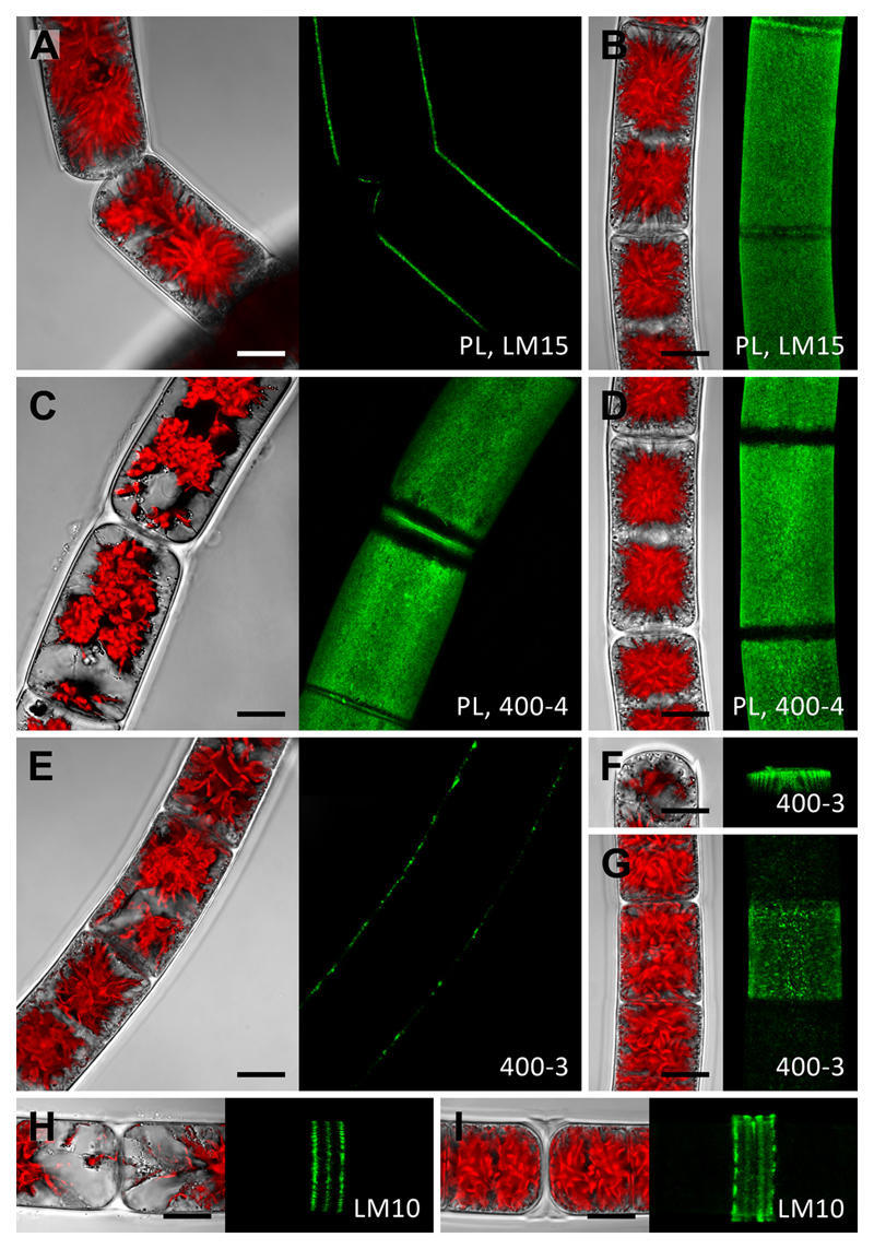
Fig. 5. Whole cell labelling of Zygnema S (CLSM micrographs).
Young (A, C, E, F, H) and old (B, D, G, I) filaments labelled with the monoclonal antibodies LM15, 400-4, 400-3 or LM10 (green). In A and E, one optical section is shown, B-D, F-I show z- projections of ~50 optical sections. The corresponding bright-field images include red chloroplast autofluorescence. (A) Detaching cells with staining in exposed cell walls. (B) Staining in outer and cross cell walls but not in ribbon-like zones close to cross cell walls. (C) Similar pattern like in (B). (D) Staining in outer cell walls. (E) Filament with patchy labelling in outer cell walls. (F) Circular staining underneath expanded terminal cross wall. (G) Central cell showing patchy straining, which is weak in adjacent cells. (H) H-shaped cell wall structure with staining in three distinct rings. (I) Prominent H-shaped cell wall structure with strong staining. Bars = 10 µm.

