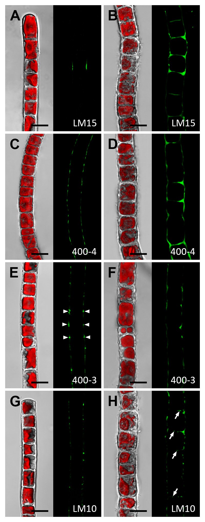
Fig. 6. Whole cell labelling of Klebsormidium crenulatum (CLSM micrographs).
Young (A, C, E, G) and old (B, D, G, H) filaments labelled with the monoclonal antibodies LM15, 400-4, 400-3 or LM10 (green). The corresponding bright-field images include red chloroplast autofluorescence. (A) Filament with weak staining in restricted areas (arrows). (B) Intense labelling in thickened cell corners between individual cells and some staining in cross cell walls. (C) Staining in outer cell walls. (D) Intense staining in cell corners and thickened cross cell walls (arrow). (E, F) Staining in cell corners (arrowheads) and occasionally in longitudinal cell walls of longer cells. (G) Punctuate staining pattern in outer cell walls. (H) Similar appearance as in (G); additionally, cross cell walls show staining (arrows). Bars = 10 µm.

