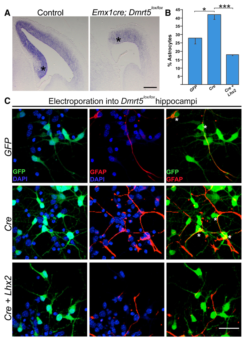
Figure 4. Lhx2 can rescue the enhanced astrogliogenesis resulting from loss of Dmrt5.
A, Lhx2 expression is decreased in EmxCre; Dmrt5lox/lox brains compared with controls. B, C, E15 embryonic hippocampi from Dmrt5lox/lox embryos were electroporated ex vivo with constructs encoding GFP, CreGFP, and CreGFP+Lhx2. After 5 d in vitro, the percentage of electroporated (GFP-expressing, green) cells that also expressed astrocyte marker GFAP (red) was scored. B, Quantification of the results shows 28% of the cells to be astrocytes upon control GFP electroporation, which increased to 42% upon loss of Dmrt5 as a result of CreGFP electroporation, and decreased to 18% upon coelectroporation of Lhx2 together with CreGFP. C, Individual GFP (green) + DAPI (blue) and GFAP (red)+ DAPI (blue) channels, as well as GFP–GFAP overlays. DAPI staining (blue) identifies the nuclei of all the cells in the field. Asterisks indicate GFP–GFAP-coexpressing astrocytes. Scale bars: A, 500 μm; C, 30 μm. *p < 0.05, ***p < 0.0001.

