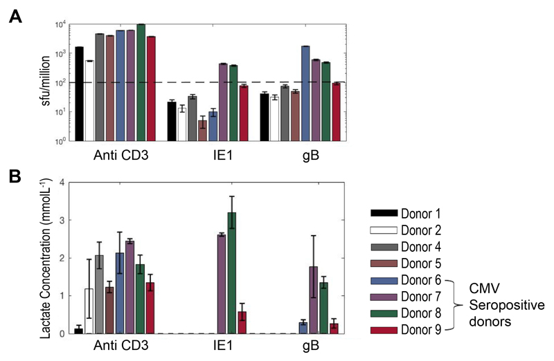
Figure 6. Screening of blood donors for responses to CMV peptides.
Data from PBMC of 8 healthy donors, cultured with soluble anti-CD3 or one of 2 different CMV peptide pools (IE1 and gB) at 1ug/mL. PBMC were plated at 105 cells per well in triplicates in 96 well plates. (A) IFNγ Elispot was performed at 48 hours and a mean sfu/million >100 was taken as a positive responder. Extracellular lactate was measured in cell culture supernatants at day 2 and day 5; (B) shows day 5 positive total lactate concentrations. All 4 seropositive donors were identified as responders to one, or both peptides by both methods.

