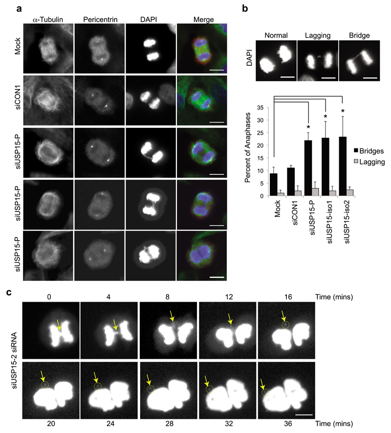
Figure 3. USP15 depletion causes anaphase bridges that can lead to micronuclei formation.
a-b, USP15 depletion in A549 cells significantly increases the number of anaphase chromosome bridges but not the number of lagging chromosomes. (a) Representative examples of anaphases observed in A549 cells, showing chromosome bridges in USP15 depleted cells. (b) Examples of each phenotype (top) with quantification of their prevalence below, data show mean of 3 independent experiments with >50 anaphases counted per condition per experiment (error bars SD, one-way ANOVA with Dunnett's multiple comparison test *P≤0.05). c, Live cell imaging demonstrates that anaphase chromosome bridges (arrow) can resolve into micronuclei (circled) in USP15 depleted cells; the timecourse begins at metaphase and extends beyond cytokinesis. All scale bars are 10μm.

