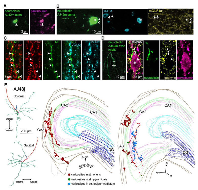
Figure 3. Teevra neurons are GABAergic, innervate PV+ neurons in the septum and target the hippocampal CA3 region.
(A, B) Neurobiotin labeled Teevra neuron, AJ42m was PV+ (magenta, main axon), SATB1+ (cyan, asterisk, nucleus) and immunonegative for mGluR1a in the plasma membrane (yellow) showing only a weak cytoplasmic signal. Note, mGluR1a+ somatic membrane labeling (yellow, arrows) of a neighboring SATB1+ cell. (C) Axon terminals of AJ42m (green) were VGAT+ (red, arrows) and VGlut2- (cyan, arrows). (D) Main axon and local terminals of AJ42m in the MS (green, box) innervating a PV+ soma (magenta, asterisk) in a basket-like formation. Note, gephyrin puncta (yellow, arrows) outlining the somato-dendritic membrane. (E) Left, reconstruction of Teevra cell AJ48j in coronal and sagittal views showing a complete ovoid dendritic field (green), soma and main axon (red) and a local axonal branch with boutons (blue). Right, the axonal varicosities (100x objective, color coded by layer) of AJ48j in two series of consecutive 80 µm thick sections (left, 4 sections; right, 3 sections), showing preferential termination in part of CA3. Coronal sections are rotated to highlight laminar selectivity (D, dorsal; M, medial, R. rostral). Imaging details (z-thickness in µm, single optical slices unless z-projection type stated): (A) 0.60 µm (B) 0.32 µm (C) 0.70 µm, maximum intensity projection (D, left) 33.60 µm, maximum intensity projection (D, right) 0.37 µm.

