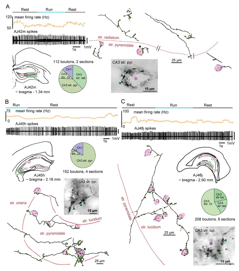
Figure 4. Teevra neurons innervate interneurons in spatially restricted domains of CA3.
Teevra neurons were identified on the bases of not changing their mean firing rate during REST vs RUN and burst duration during CA1 theta (upper panels). Reconstructions of axonal collaterals (green, boutons) of labeled cells reveal that Teevra neurons, AJ42m (A), AJ45h (B) and AJ48j (C) innervate interneurons in the CA3 region. Innervated interneuron somata (shaded pink) are identified by endogenous biotin in mitochondria. Pie charts show representative samples of bouton distribution in different areas and layers. Light micrographs of axonal varicosities (arrows) visualized by HRP enzyme reaction adjacent to cell bodies of individual interneurons (asterisks) rich in endogenous biotin in mitochondria (black in cytoplasm) as revealed by the color reaction.

