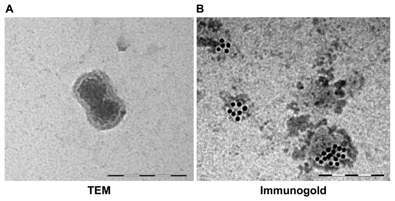
Figure 3. Size, morphology and expression profile characterize exosomes.
(A) Transmission electron microscopy (TEM) of MRC5 fibroblast exosome (120 000x) demonstrating bi-layered structure. (B) Immunogold staining of primary mesenchymal stem cell exosomes with CD63. Scale bars in both panels represent 200 nm.

