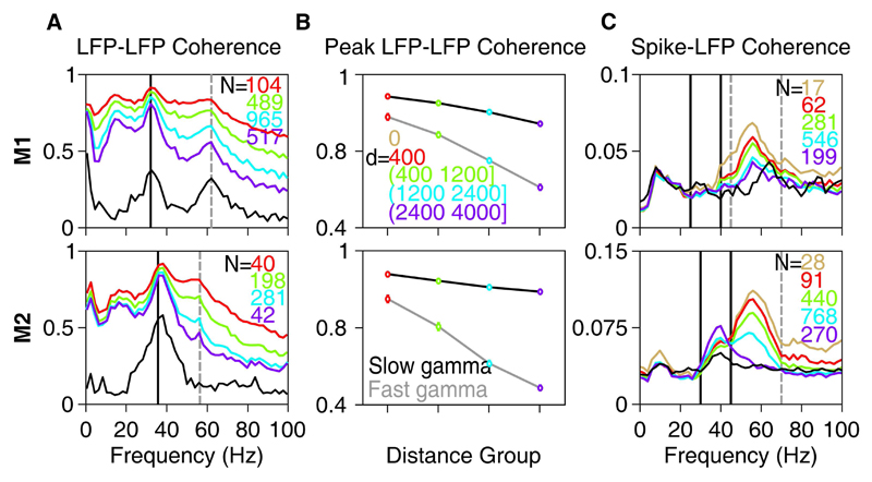
Figure 7. Field-field and Spike-field Coherence.
(A) LFP-LFP phase coherence spectra for different inter-electrode distances for Monkeys 1 (top row) and 2 (bottom row). Inter-electrode distance ranges (d, in µm) are mentioned in B. The number of pairs (N) for each group is indicated on the top right corner. LFP-LFP phase coherence when both are taken from the same electrode (i.e., inter-electrode distance of zero) is trivially 1 at all frequencies and is therefore omitted. Mean LFP-EEG phase coherence is shown in black. (B) Average LFP-LFP phase coherence at the peak slow (32 and 36 Hz for the two monkeys) and fast gamma bands (62 and 56 Hz) as a function of inter-electrode distance. (C) Mean Spike-LFP coherence for spike-LFP pairs separated by different inter-electrode distances. Mean Spike-EEG coherence is shown in black.

