Figure 21.
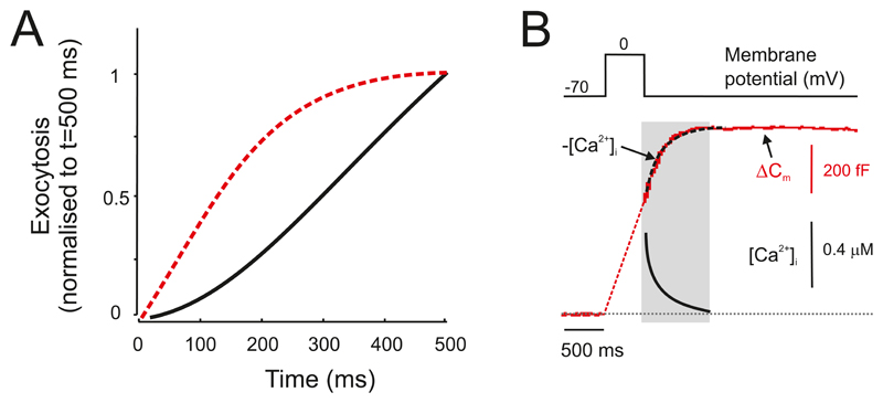
A: Relationship between exocytosis (measured as the depolarization-evoked increase in membrane capacitance) in isolated human β-cells (solid black line) and isolated mouse β-cells (dashed red line). Responses have been normalized to exocytosis elicited by a 500 ms depolarization. Note that depolarizations <100 ms evoke only small increases in capacitance in human β-cells. In β-cells within intact human islets there is an additional, rapid component of exocytosis (568). Measurements were performed in the presence of 0.1 mM cAMP. Modified from (90, 170). B: Example of capacitance increase (ΔCm, red trace) elicited by a 500-ms depolarization in an isolated human β-cell. Note that in human β-cells, unlike mouse β-cells (Figure 19B), exocytosis continues for ~500 ms beyond the end of the pulse (grey rectangle). The dashed red line represents a linear extrapolation of the post-depolarization exocytosis to the start of the depolarization. The blue trace shows the decay of depolarization-evoked [Ca2+]i recorded in a β-cell following membrane repolarization. The dashed line superimposed on the capacitance trace is the inverted [Ca2+]i signal. Modified from (90, 563). This type of ‘asynchronous’ exocytosis suggest that exocytosis in human β-cells is determined by the bulk rather than the local (near-membrane) [Ca2+]i.

