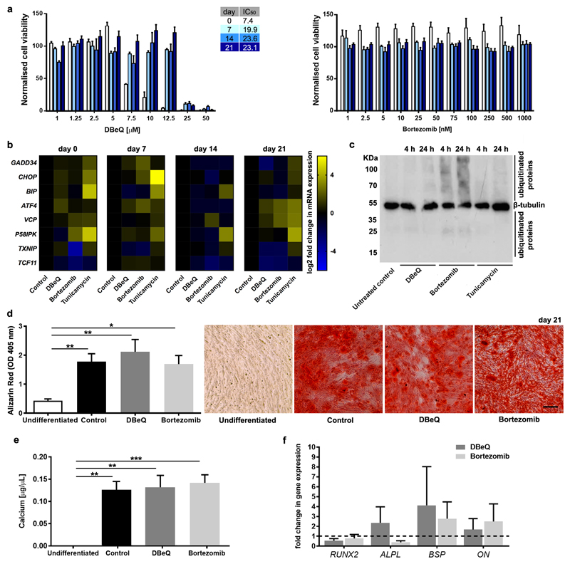
Fig. 1. Mild proteostasis perturbations do not grossly affect the osteogenic differentiation of hMSC.
a, Normalised viability of hMSC undergoing osteogenic differentiation treated on days 0, 7, 14, and 21 with the indicated concentrations of DBeQ or bortezomib for 48h (n = 3). b, Heatmaps showing relative mRNA expression levels (normalised to undifferentiated controls) for GADD34, CHOP, BIP, ATF4, VCP, P58IPK, TXNIP and TCF11 in differentiating hMSC on days 0, 7, 14, and 21 after treatment for 24 h with DBeQ (5 μM), bortezomib (20 nM), or tunicamycin (5 μg/mL). c, Immunoblotting for β-tubulin and ubiquitinated proteins on whole cell extracts from undifferentiated hMSC untreated (control) or treated for 4 h or 24h with 5 μM DBeQ, 20 nM bortezomib, or 5 μg/mL tunicamycin. d, Quantification and representative micrographs showing Alizarin Red S staining (Scale bar = 200µm) and e, colorimetric calcium content quantitation of differentiated hMSC cultures. f, relative mRNA expression levels for markers of osteogenic differentiation compared to undifferentiated controls, which are set to 1 (dotted horizontal line). In a, d-f, plots show mean + SEM and in b, heat maps show mean of log 2 fold changes in gene expression (normalised to undifferentiated controls) for hMSC from 3 different donors. In d-f, a Kruskal-Wallis non-parametric test followed by Dunn’s Multiple Comparison test was used to detect statistical significance, *p < 0.05, **p < 0.01 and ***p < 0.001. For detailed n and p values see Supplementary Tables 1-4.

