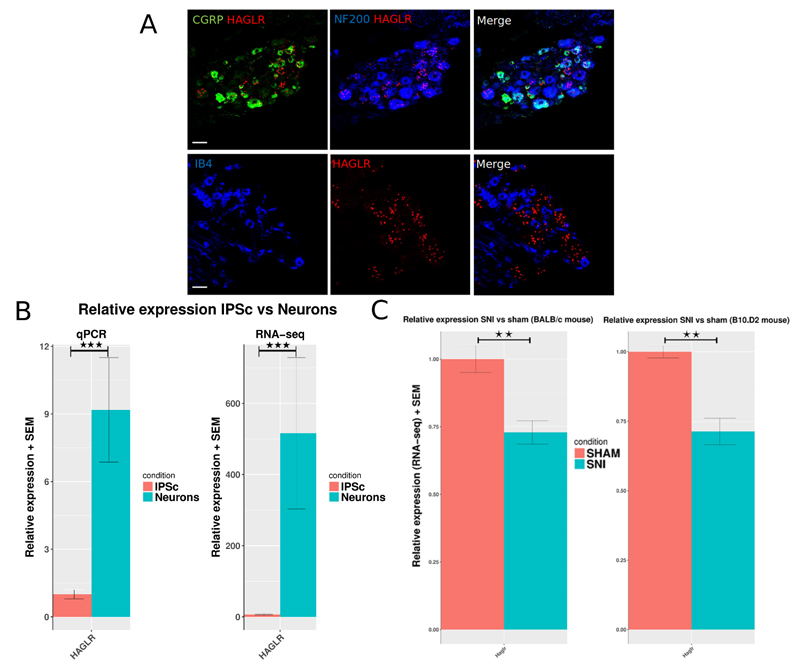
Figure 5.
A: In-situ hybridisation for Haglr LncRNA (mouse ortholog of human HAGLR) shows expression in mouse WT L4 DRG. The ISH signal was developed using a fast red reaction. From right to left: Representative images of mouse DRG sections stained for Haglr (red) and NF200 (blue), CGRP (green), and IB4 (blue). Scale bar 50µm. B: Quantification of expression change of HAGLR in human IPSC vs IPSC-derived neurons. Relative expression assessed by qPCR of HAGLR LncRNA. C: RNA-seq determined relative expression in SNI vs Sham BALB/c and B10.D2 mice DRG. Data is presented as mean plus SEM. P < 0.5 *, p < 0.01 **, P < 0.001 ***.

